Medical History Shattered in Sri Lanka: Surgeons Extract Record-Breaking 3.1-Kilogram Bladder Stone in Remarkable Operation

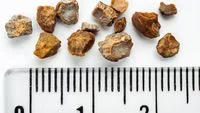
A team of specialized medical professionals in Sri Lanka has successfully performed a historic and highly complex surgical procedure, removing an extraordinarily massive urinary bladder stone from a 55-year-old male patient. The calcified mass, weighing an astonishing 3.1 kilograms (6.8 pounds) and measuring approximately 17 centimeters (6.7 inches) in length, has stunned the global medical community.
Conducted at the Trincomalee District General Hospital, the successful operation was spearheaded by senior consultant urologist Dr. Pragaladhan Balakrishnan and his dedicated surgical team. According to preliminary medical assessments and historical clinical comparisons, the dimensions and sheer weight of the extracted mass suggest it may be the largest urinary bladder stone ever surgically removed and formally documented in medical history. While official global verification bodies are still reviewing the case documentation, the clinical implications of the procedure highlight both the triumphs of modern urological surgery and the hidden dangers of untreated urinary tract complications.
Anatomy of a Medical Marvel: Size, Weight, and Comparisons
To fully comprehend the extraordinary nature of the Sri Lankan case, medical experts have drawn comparisons to everyday objects and official world records. Weighing 3.1 kilograms, the stone is roughly equivalent to the weight of a healthy newborn human baby. In terms of physical dimensions, measuring 17 centimeters long, the calcified mass is comparable in size to a large ostrich egg.
Prior to this intervention, the official benchmark recognized by Guinness World Records for the largest urinary bladder stone on record belonged to a patient from Brazil. In a landmark 2003 medical case, doctors extracted a bladder stone weighing 1.9 kilograms (4.2 pounds) with a length of 17.9 centimeters. While the Brazilian specimen held a slightly longer length, the mass removed in Trincomalee substantially exceeds it in overall weight by over a full kilogram.
Dr. Balakrishnan noted that hospital archives and available global medical literature indicate that this specimen stands as a record-breaking surgical removal. Nevertheless, institutional validation through international medical registries and record-keeping authorities remains pending as the surgical team compiles comprehensive documentation, imaging scans, and post-operative reports.
Chronology and Clinical Background of the Patient
The path that led the 55-year-old Sri Lankan patient to the operating table began long before the discovery of the colossal stone. Prior to his admission to the Trincomalee District General Hospital, the patient had suffered a debilitating stroke. Neurological events of this nature frequently disrupt normal bodily functions, including the intricate neural pathways that govern urinary bladder control.
During his subsequent hospitalization and stroke rehabilitation, medical staff observed that the patient was experiencing severe complications while attempting to urinate. Initial bedside assessments revealed profound urinary retention, prompting physicians to order diagnostic imaging. Ultrasonography and subsequent radiological evaluations revealed a shocking discovery: a dense, gargantuan mass occupying virtually the entire volume of the patient’s urinary bladder.
Recognizing the extreme risks associated with such a massive foreign body—ranging from total urethral obstruction to bladder rupture and fatal urosepsis—Dr. Balakrishnan and his team formulated an urgent surgical plan. The high-risk open urological procedure was executed with meticulous precision to avoid catastrophic hemorrhage or damage to surrounding pelvic organs. Following the successful extraction of the 3.1-kilogram mass, hospital spokespeople confirmed that the patient’s post-operative recovery has been remarkably smooth, with his vital signs remaining stable and his overall physical condition assessed as very good.
Pathophysiology: Understanding Urinary Bladder Stones
To understand how a mineral mass can grow to the size of an internal organ, medical science looks at the fundamental processes of crystal formation within the urinary tract. Urinary bladder stones, scientifically referred to as cystoliths, are hard masses of mineral deposits that develop when the chemical composition of urine becomes excessively concentrated.
In a healthy human body, the kidneys filter waste products from the blood, producing urine that is regularly and completely expelled from the bladder during urination. However, when the bladder fails to empty entirely, residual urine remains trapped inside. Over time, minerals such as calcium, magnesium, oxalate, and uric acid dissolved in the stagnant urine begin to separate, precipitate, and form microscopic crystals.
Under normal circumstances, these tiny crystals are flushed out of the body naturally. When urinary retention persists, however, the crystals accumulate, adhere to one another, and progressively layer over months or even years. While many small bladder stones pass unnoticed or cause mild discomfort, stones measuring greater than 4 centimeters are clinically classified as giant bladder stones. At approximately 17 centimeters, the Sri Lankan patient’s stone far surpasses this threshold, representing an extreme pathological extreme of chronic urinary stasis.
Etiology and Risk Factors: Why Do Giant Stones Form?
The development of a bladder stone of this magnitude does not occur in a vacuum; it is invariably the symptom of an underlying medical condition that severely impairs normal bladder evacuation. Medical researchers and urologists have identified several primary risk factors that exponentially increase an individual’s susceptibility to massive stone formation:
- Neurological Impairment: Conditions that disrupt the brain-bladder connection—such as strokes, spinal cord injuries, multiple sclerosis, and neurodegenerative diseases—prevent the detrusor muscle of the bladder from contracting effectively, resulting in chronic incomplete emptying.
- Bladder Outlet Obstruction: Anatomical blockages that impede the flow of urine, most notably benign prostatic hyperplasia (BPH) in aging men or urethral strictures, force the bladder to strain against resistance, often leaving behind a pool of residual urine.
- Chronic Urinary Tract Infections (UTIs): Persistent or recurrent bacterial infections alter the chemical makeup of urine, promoting the precipitation of mineral salts and encouraging stone crystallization.
- Indwelling Medical Devices: Long-term utilization of urinary catheters can introduce foreign surfaces upon which mineral encrustations rapidly form and multiply.
- Metabolic and Dietary Factors: Dehydration, diets high in specific minerals, and metabolic disorders can elevate the concentration of stone-forming crystals in the renal system.
In the case of the Sri Lankan patient, his history of a stroke served as the primary catalyst, paralyzing or severely impairing the neurological signals required for proper bladder emptying, thereby creating an ideal internal environment for undisturbed stone growth.
Clinical Symptoms and Warning Signs
Bladder stones do not always manifest with immediate or dramatic symptoms, particularly in their early stages. Small stones may simply bounce harmlessly against the bladder wall or pass unassisted through the urethra. However, as a stone grows—let alone reaches record-breaking proportions—it inevitably triggers a cascade of distressing and painful clinical symptoms.
Medical professionals advise individuals to monitor for several hallmark warning signs associated with bladder stones:
- Dysuria: Experiencing sharp pain, discomfort, or a burning sensation during the act of urination.
- Obstructive Voiding: Difficulty initiating a urine stream, or experiencing a stream that starts and stops intermittently without warning.
- Weak Urinary Flow: A noticeable reduction in the force, volume, and projection of the urinary stream.
- Frequency and Urgency: A sudden, overwhelming compulsion to urinate frequently, often yielding only small amounts of urine.
- Hematuria: The presence of visible blood in the urine, caused by the abrasive physical friction of the stone rubbing against the delicate mucosal lining of the bladder wall.
- Suprapubic Pain: A dull, persistent ache or pressure localized in the lower abdomen, directly above the pubic bone.
- Complete Urinary Retention: The total inability to void urine, constituting an immediate medical emergency.
Healthcare providers emphasize that while these symptoms do not exclusively point to bladder stones—often overlapping with prostate conditions or urinary infections—their persistence necessitates thorough urological evaluation via cystoscopy, ultrasound, or computed tomography (CT) imaging.
Broader Implications and Public Health Lessons
The removal of the 3.1-kilogram bladder stone in Trincomalee transcends the boundaries of a mere medical curiosity; it serves as a stark reminder of the critical importance of proactive healthcare, routine medical monitoring, and the prompt management of urinary symptoms.
Urologists worldwide point out that bladder stones rarely attain massive proportions if underlying voiding dysfunctions are diagnosed and addressed early in their progression. Modern urology offers numerous minimally invasive interventions—such as cystolitholaprese, laser lithotripsy, and endoscopic stone extraction—that can safely eliminate stones long before open surgical intervention becomes mandatory.
Furthermore, the case underscores the vulnerability of vulnerable patient populations, such as stroke survivors and elderly individuals with mobility or cognitive limitations. Caregivers, family members, and primary healthcare providers must remain vigilant regarding changes in urinary habits, bathroom frequency, or signs of discomfort in patients who may be unable to articulate their symptoms independently.
Beyond treating the physical stone itself, medical consensus dictates that comprehensive long-term management must focus on eradicating the root cause of urinary stasis. Whether through pharmacological management of prostate enlargement, physical therapy for neurological deficits, or the adjustment of catheterization protocols, resolving the underlying pathology is the only reliable method to prevent the recurrence of stone formation.
As the global medical community awaits the final administrative verification from official record-keeping organizations regarding the Sri Lankan case, the extraordinary procedure stands as a testament to the skill of the surgical team at Trincomalee District General Hospital and a profound cautionary tale about the silent dangers of neglected urological health.
Related posts: