Understanding Bone Metastasis and the Advanced Role of SPECT-CT Imaging in Modern Oncology Care

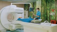
The silent progression of cancer to the skeletal system remains one of the most challenging hurdles in modern oncology, particularly in prevalent malignancies such as breast and prostate cancers. Often developing without immediate, noticeable symptoms in its earliest stages, bone metastasis can severely compromise a patient’s structural integrity, mobility, and overall quality of life. To combat this hidden threat, advancements in medical imaging have shifted the paradigm of early detection and risk evaluation. Among these innovations, Single-Photon Emission Computed Tomography combined with Computed Tomography, commonly known as SPECT-CT, has emerged as a cornerstone technology. By merging functional cellular data with precise anatomical mapping, this sophisticated imaging tool empowers physicians to detect abnormal bone activity, pinpoint metastatic lesions with remarkable accuracy, and formulate targeted treatment strategies tailored to the unique physiological landscape of each patient.
The Clinical Challenge of Bone Metastasis
Malignant tumors do not always remain confined to their primary site of origin. Through a complex biological cascade known as metastasis, cancer cells can detach from the primary tumor, invade blood vessels or lymphatic channels, and travel to distant organs. The skeletal system is one of the most frequent destinations for metastatic cells, particularly in cancers of the breast, prostate, lung, and kidney.
When cancer cells infiltrate the bone matrix, they disrupt the delicate balance between bone-forming cells (osteoblasts) and bone-resorbing cells (osteoclasts). This disruption leads to structural weakening, leaving the skeletal system vulnerable to a cascade of debilitating complications. Patients frequently suffer from severe, persistent bone pain, pathological fractures resulting from minimal or no trauma, hypercalcemia, and spinal cord compression—a true medical emergency that can lead to permanent paralysis if left unaddressed.
Historically, detecting these changes in their infancy was difficult. Conventional X-rays often fail to reveal metastatic lesions until a significant portion of the bone mineral content has already been destroyed. While traditional bone scans offer high sensitivity, they frequently lack specificity, making it challenging to differentiate between benign conditions—such as fractures, arthritis, or degenerative joint disease—and true malignant metastasis. This diagnostic grey area has long driven the medical community to seek more precise, high-resolution diagnostic modalities.
The Technological Leap: How SPECT-CT Works
The integration of Single-Photon Emission Computed Tomography (SPECT) and Computed Tomography (CT) into a single hybrid imaging session represents a monumental leap forward in diagnostic radiology and nuclear medicine.
To understand the value of this technology, one must examine its dual components. SPECT imaging relies on the administration of a safe, radioactive tracer that travels through the bloodstream and accumulates in areas of active bone remodeling or increased cellular turnover. This functional imaging highlights "hot spots" where physiological changes are occurring, even before structural damage becomes visible. However, SPECT alone often falls short in providing precise anatomical localization.
This is where the CT component becomes indispensable. By capturing high-resolution cross-sectional X-ray images of the skeletal anatomy during the same scanning session, the hybrid system overlays the functional metabolic data from the SPECT scan onto the anatomical roadmap provided by the CT scan. The result is a multi-dimensional, comprehensive visualization that allows clinicians to see not only that an abnormality exists, but precisely where it is located, how large the affected area is, and how it interacts with surrounding tissues, nerves, and joints.
Clinical Perspectives and Expert Insights
Medical specialists emphasize that managing advanced oncological cases requires deep, granular insight into the precise extent of skeletal involvement. Dr. Yustia Tuti, Sp.KN-TM, Subsp.Onk (K), a nuclear medicine specialist at Mayapada Hospital Jakarta Selatan, highlights the clinical necessity of comprehensive bone evaluations.
"Pada pasien dengan metastasis tulang, kita perlu melihat bukan hanya apakah terdapat lesi, yaitu area tulang yang menunjukkan kelainan, tetapi juga di mana lokasi penyebaran sel kanker, seberapa luas, dan bagaimana kondisi tulang yang terdampak," explained Dr. Yustia, emphasizing that understanding the exact topography of the disease is vital for anticipating and mitigating catastrophic skeletal-related events.
Without this detailed mapping, predicting complications such as pathological fractures or neurological compromise remains speculative. By leveraging SPECT-CT, multidisciplinary medical teams can evaluate the structural integrity of weight-bearing bones, assess the risk of spinal cord impingement, and intervene proactively before irreversible damage occurs.
Furthermore, distinguishing between true metastasis and benign bone pathologies is crucial to avoid subjecting cancer survivors to unnecessary anxiety, misdiagnoses, or inappropriate therapeutic adjustments. Dr. Andreas Lim, Sp.KN-TM, Subsp. Onk. (K), FANMB, also a nuclear medicine specialist at Mayapada Hospital Jakarta Selatan, underscores the diagnostic clarity that hybrid imaging provides.
"Dengan melihat hasil pemeriksaan SPECT-CT, dokter dapat menilai apakah temuan tersebut lebih mengarah pada metastasis atau perubahan lain pada tulang. Pemeriksaan ini juga dapat membantu memperjelas temuan yang masih meragukan, sehingga langkah evaluasi selanjutnya dapat ditentukan sesuai kondisi pasien," noted Dr. Andreas Lim. This diagnostic precision ensures that oncologists and surgeons can design treatment pathways based on absolute clarity rather than ambiguity.
Streamlining Workflow and Enhancing Treatment Efficiency
In the fast-paced environment of modern healthcare, efficiency directly correlates with patient outcomes. The deployment of advanced imaging modalities like SPECT-CT streamlines the clinical workflow for multidisciplinary oncology teams. By delivering definitive answers in a single examination, the technology minimizes the need for redundant, sequential imaging tests, thereby reducing patient exposure to radiation, lowering overall diagnostic costs, and accelerating the timeline from diagnosis to intervention.
When oncologists, radiologists, orthopedic surgeons, and nuclear medicine physicians review a comprehensive whole-body SPECT-CT scan, they are equipped with a unified dataset. This shared understanding facilitates more efficient tumor board discussions, where treatment protocols—ranging from systemic chemotherapy and targeted hormone therapies to localized radiation therapy and prophylactic orthopedic stabilization—are finalized with pinpoint accuracy.
The Evolution of Comprehensive Cancer Care in Indonesia
Access to state-of-the-art diagnostic tools has historically been concentrated in a handful of global medical hubs, forcing patients to travel abroad for advanced oncological evaluations. However, recent healthcare infrastructure developments within Indonesia have significantly narrowed this gap, bringing world-class medical technology directly to domestic patients.
A prime example of this domestic advancement is the Nuclear Medicine Polyclinic at Mayapada Hospital Jakarta Selatan (MHJS). Equipped to perform comprehensive whole-body imaging according to individual patient clinical requirements, the facility integrates advanced nuclear medicine diagnostics into a broader, cohesive healthcare framework known as Comprehensive Cancer Care.
Within this integrated model, diagnostic imaging does not exist in a vacuum. Instead, it serves as the foundational pillar of the hospital’s Oncology Center. This specialized ecosystem relies on close collaboration among multidisciplinary medical teams who convene regularly within a structured Tumor Board. These medical boards analyze complex cases collectively, ensuring that every therapeutic decision—from initial staging to palliative supportive care—is evidence-based, personalized, and executed with optimal timing.
Moreover, recognizing the psychological and logistical burdens faced by cancer patients and their families, institutions like Mayapada Hospital incorporate dedicated Patient Navigators into their care models. These professionals act as compassionate guides, assisting patients through the complex maze of hospital appointments, diagnostic procedures, treatment cycles, and follow-up care.
Broad Implications for Public Health and Future Outlook
The broader implications of integrating advanced hybrid imaging into standard oncology protocols extend far beyond individual hospital walls. As cancer incidence rates continue to rise globally, driven by demographic shifts, aging populations, and lifestyle factors, the demand for precise, early diagnostic tools will only intensify.
Early detection of bone metastasis not only preserves functional independence and alleviates chronic pain for patients, but it also alters the economic trajectory of long-term cancer care. By preventing catastrophic skeletal complications—such as sudden pathological fractures requiring emergency orthopedic surgeries and prolonged hospitalizations—advanced imaging ultimately reduces the cumulative burden on healthcare systems.
Patients and families seeking more information regarding advanced diagnostic services, specialist consultations, or comprehensive oncological evaluations can access support through dedicated institutional communication channels. Facilities such as Mayapada Hospital provide accessible coordination via their central Call Center at 150770, direct messaging via WhatsApp at 0817-1715-077, or the 24-hour Emergency hotline at 150990. Additionally, digital health tools, such as the MyCare mobile application, offer integrated platforms for patients to manage their appointments and monitor personal health metrics—including heart rate, daily step counts, caloric expenditure, and Body Mass Index (BMI)—fostering a holistic approach to personal wellness and disease prevention in the modern digital age.